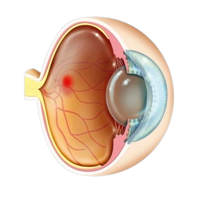
RETINA
Did you know that your clear view of the world, the one that allows you to read, drive and recognize faces, relies on the health of your retinas?
So, what is the retina and how does it help us see?
How We See
Think of your eye as a camera. Light enters through the cornea and is controlled by the iris and pupil, which function like a camera shutter, opening (dilating) and closing to allow more light in dark conditions and less light in bright conditions.
This focused light is directed onto the retina, a thin layer of light-sensitive nerve tissue that lines the back of the eye, like film in a camera. Images are focused at its center (known as the macula) and converted into electrical impulses that are carried to the brain by the optic nerve, where they are translated into sight!
Maintaining Retina Health for Good Vision
Retinal disease is a leading cause of blindness and early detection is the game changer.
Here’s how to safeguard your sight:
- Be aware of your risk factors, which may include age, family history or pre-existing health conditions.
- Pay attention to visual changes and visit an eye physician right away if experience symptoms such as blurry or distorted vision, if straight lines appear wavy, or you see dark spot, flashes of light or floaters.
- Have a regular dilated retina exam.
- See a retina specialist for expert care of retinal diseases/conditions.
Problems Related to Retina

A detached retina is when the retina lifts away from the back of the eye. The retina does not work when it is detached, making vision blurry. A detached retina is a serious problem. An ophthalmologist needs to check it out right away, or you could lose sight in that eye.
How do you get a detached retina?
As we get older, the vitreous in our eyes starts to shrink and get thinner. As the eye moves, the vitreous moves around on the retina without causing problems. But sometimes the vitreous may stick to the retina and pull hard enough to tear it. When that happens, fluid can pass through the tear and lift (detach) the retina.
Who is at risk for a detached How is a detached retina retina?
- You are more likely to have a detached retina if you:
- Need glasses to see far away (are nearsighted)
- Have had cataract, glaucoma, or other eye surgery
- Take glaucoma medications that make the pupil small (like pilocarpine)
- Had a serious eye injury
- Had a retinal tear or detachment in your other eye
- Have family members who had retinal detachment
- Have weak areas in your retina (seen by an eye doctor during an exam)
Early signs of a detached retina
A detached retina has to be examined by an ophthalmologist right away. Otherwise, you could lose vision in that eye. Call an ophthalmologist immediately if you have any of these symptoms:
- Seeing flashing lights all of a sudden. Some people say this is like seeing stars after being hit in the eye.
- Noticing many new floaters at once. These can look like specks, lines or cobwebs in your field of vision.
- A shadow appearing in your peripheral (side) vision.
- A gray curtain covering part of your field of vision.

Macular hole is when a tear or opening forms in your macula. As the hole forms, things in your central vision will look blurry, wavy or distorted. As the hole grows, a dark or blind spot appears in your central vision. A macular hole does not affect your peripheral (side) vision.
What causes a macular hole?
Age is the most common cause of macular hole. As you get older, the vitreous begins to shrink and pull away from the retina. Usually the vitreous pulls away with no problems. But sometimes the vitreous can stick to the retina. This causes the macula to stretch and a hole to form.
Sometimes a macular hole can form when the macula swells from other eye disease. Or it can be caused by an eye injury.
How is a macular hole diagnosed?
Get more information about macular hole from EyeSmart—provided by the American Academy of Ophthalmology—at aao.org/macular-hole-link.
